|
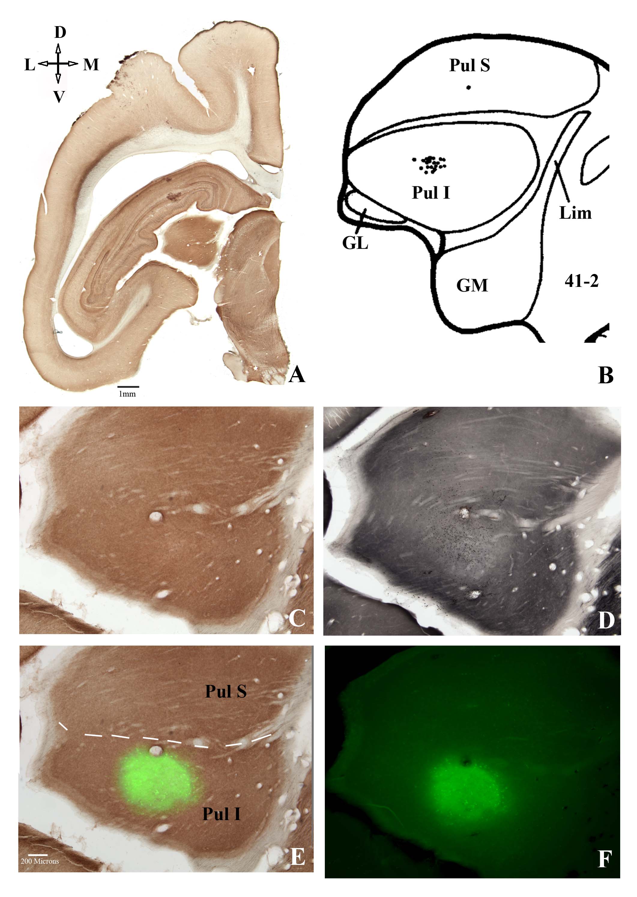
| Fig. 1 shows the location of a restricted blocking injection of muscimol in bush baby inferior pulvinar (PulI). A AChE stained coronal section through the divisions of pulvinar. B Retrogradely labeled cells in bush baby following a restricted injection near the V1/V2 border from Raczkowski & Diamond (150) . C and D Two adjacent sections of the portion of PulIwe mapped electrophysiologically and injected 200nl of muscimol/dextran and subsequently stained for AChE and CO, respectively. Note that in the CO section the area injected shows a reduction in CO activity which corresponds exactly to the injected zone shown in the original fluorescent image in F and the double exposure image in E. The dotted line in E divides superior (pulS) from inferior pulvinar (pulI). Other abbreviations: D, dorsal; M, medial; L, lateral; V, ventral; GL, lateral geniculate nucleus; GM, medial geniculate nucleus; Lim, limitans; 41-2, section number in Raczkowski & Diamond (150) . |