Casagrande
Vision Research Lab
Lab Activities
2008 Lab Poster (pdf)
2007 Lab Poster (pdf)
|
 Scientific Research Scientific Research |
Lab Meeting
|
 Beer & Paper (Lab Seminar) Beer & Paper (Lab Seminar)
|
|
Drawing by David Royal |
Weekly lab meetings are usually held on Friday mornings. The meetings
are important for lab members to exchange information, solve problems and
general new ideas. Don't be late! Otherwise, you know what...
(ask Julie) :) |
This is an informal lab research seminar, but it is an important occasion. We take turns leading the seminar, including selecting research papers of interest to us and buying beer and snacks. We also practice our talks and comment on our own papers. |
|
|
|
|
|
|
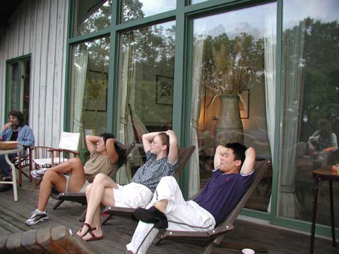
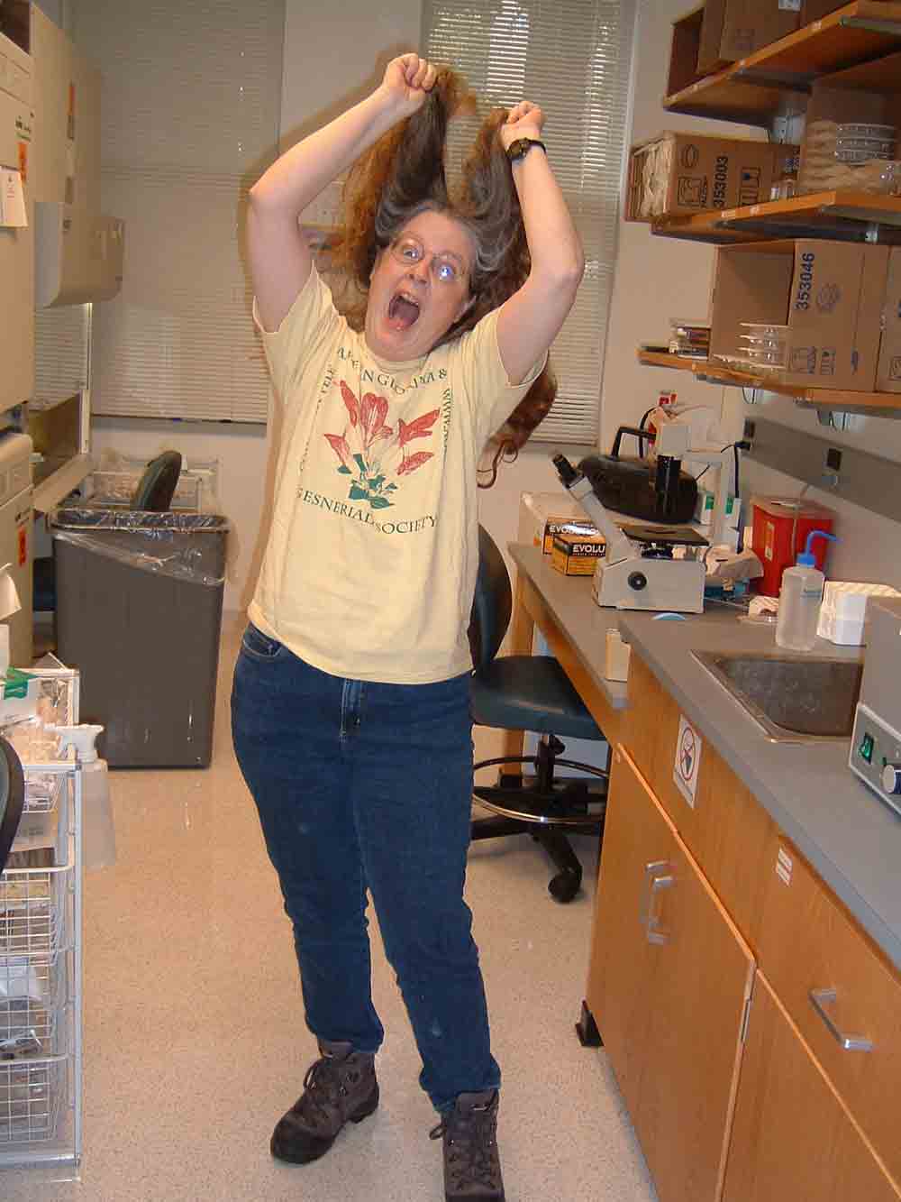
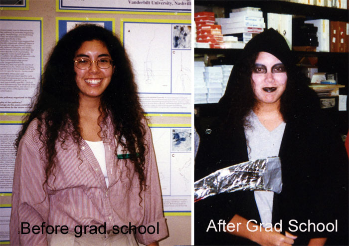
 Lab Socials Lab Socials
If you stay in our lab for a while, you will notice that it is a lively and fun lab, always filled with joy, music and laughter... See some pictures
|
|
|
 Research
Projects Research
Projects |
Major
Investigator(s) |
Project
Description |
|
(1) Electrophysiology
in behavior-trained awake monkeys
NSF Related Work
|
|
The lateral geniculate nucleus (LGN) may do more then simply
relay sensory signals since it receives numerous extraretinal inputs that
together far out-number, in terms of synapses, retinal input. The LGN also
contains a complex set of circuits involving inhibitory interneurons. We
hypothesize that LGN neurons are part of a dynamic network which carries
information relevant not only to sensory quality, but also to behavioral
state including signals related to visual attention, task relevance, and
eye movements. We are testing this hypothesis and examining how information
is regulated at the level of the LGN using standard electrophysiological
techniques in awake behaving macaque monkeys. |
|
| (2) Studies
of functional organization of visual cortex by using optical imaging techniques |
|
To investigate how neurons in visual cortex are functionally organized
to process different aspects of visual information, and examine the relationships
between functional maps or modules. A). How are basic receptive field properties
(such as orientation preference, ocular dominance and spatial frequency
selectivity) of V1, V2 and V3 organized in bush babies and owl monkeys? B). Do V1 CO compartments
encode segregated visual information? Does V2 and V3 have distinct functional
compartments to separately process luminance difference, motion or forms
and textures? |
|
| (3) Feedback pathways from
V1 to the LGN in the primate visual system |
|
Information traveling to visual cortex has a parallel organization
that is seen in many mammalian species. In primates these parallel
streams of information are segregated in to the magnocellular (M), parvocellular
(P) and koniocellular (K) pathways. One of the goals of this research
is to determine how these pathways are reflected in the organization of
corticogeniculate feedback from primary visual cortex (V1) to the lateral
geniculate nucleus (LGN) in the thalamus. |
|
| (4) Receptive field properties of parallel
visual pathways (LGN cells) |
Xiangmin Xu (currently Assistant Professor, Department of Anatomy & Neurobiology, University of California, Irvine)
Paper (PDF)
|
By analogy to previous work on lateral geniculate nucleus (LGN) magnocellular
(M) and parvocellular (P) cells our goal was to construct a physiological
profile of koniocellular (K) cells that might be linked to particular visual
perceptual attributes. Our findings suggest that the K cells consist
of several classes, some of which could contribute to conventional aspects
of spatial and temporal resolution. |
|
| (6) Metabotropic glutamate receptor 5 (mglur5) shows different patterns of localization within the parallel visual pathways in primates |
Yura Shostak (now in Belarus)
Paper
Currently need new researchers to carry on this project. Please join us!
|
Objectives:
1) To localize mGluR5 receptors within the layers of V1 in relation
to the K, M, and P LGN pathways.
2) To compare the mGluR5 synaptic localization and distribution in
macaque monkey V1 with that in squirrel monkey.
|
|
| (7) Immunochemical studies: the distribution of koniocellular lateral geniculate nucleus (LGN) cells in macaque monkey |
Currently need new researchers to carry on this project. Please join us!
|
To estimate the distribution of K LGN cells we immunostained sections to reveal K cells using anti-alphaCaMII kinase. K cell density was measured at three eccentricities according to the map of Malpeli et al. (1996): 0-5 deg (fovea & parafoveal), 5-10 deg (paracentral), 10-17 deg (peripheral). Results show that the average density of K cells in these areas is 143.3+38.1 cells/mm2, 100.5+28.6 cells/mm2 and 74.2+14.9 cells/mm2, respectively. Extending this analysis to estimate the number of K cells per unit area of visual space should indicate whether the density of K cells is sufficient to support the chromatic acuity reported for separate M/L and S cone channels. |
|
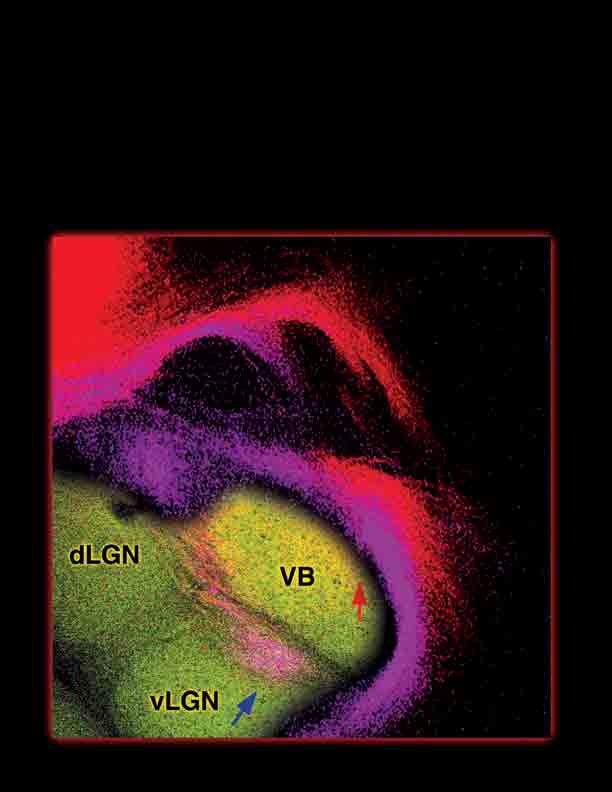
(8) Molecular aspects of visual system development and
orgnization:
Cortical development in mutant mice lacking polysialylated
neural cell adhesion
molecule
|
|
Cell adhesion molecules are proposed to mediate contact-dependent axon
guidance events. Axon guidance involves a number of elements including
axon outgrowth, degree of fasciculation,
targeting, and branching. We have used mice lacking L1, NCAM-180
(neural cell adhesion molecule-180), or polysialic
acid (PSA), to investigate the role of these molecules in the process
of thalamocortical development and axon pathfinding. |
|
|
|
|
|